Understanding Cervical Polyps
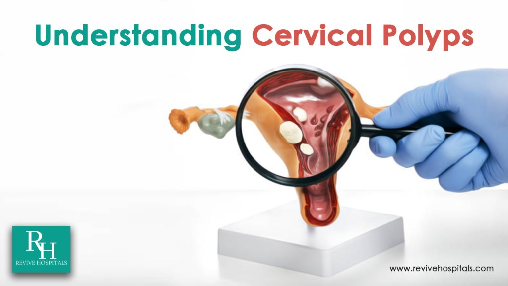
Cervical polyps are a common gynecological condition that affects women any age group. They appear as fingerlike growths on the uterus’s lower part, which connects with the vagina. They are not harmful but they can cause discomfort and irregular bleeding. This condition may occur with an abnormal response to increased levels of the female hormone estrogen, Chronic inflammation, and Clogged blood vessels in the cervix. Diagnosing and managing cervical polyps involve various techniques, and some of them are hysteroscopy and ultrasound. Let’s get a better look into these procedures
The Uses of Hysteroscopy
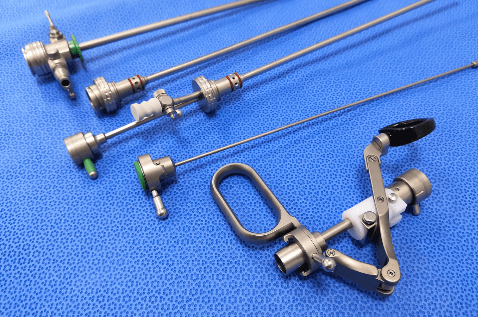
Hysteroscopy is a procedure performed by surgeons to get a better look inside the uterus to examine abnormal bleeding. It involves the insertion of a thin, lighted tube with a camera, called a hysteroscope, into the uterus through the cervix. It allows doctors to directly visualize the uterine cavity and diagnose and treat various conditions, including cervical polyps.
Here are some key uses of hysteroscopy:
Diagnosis: Hysteroscopy is a very effective procedure used in diagnosing cervical polyps, as it provides a clear and detailed view of the uterine lining and the polyp itself. This method helps differentiate between polyps and other uterine abnormalities or growths.
Removal (Polypectomy): If a cervical polyp is identified during hysteroscopy, it can often be removed during the same procedure. A small instrument is inserted through the hysteroscope to grasp and remove the polyp. The removal of the polyp is called Hysteroscopy Polypectomy.
Examination of Abnormal Bleeding: Hysteroscopy can help diagnose the cause of abnormal uterine bleeding, heavy menstrual bleeding, and irregular spotting, including bleeding caused by cervical polyps. Doctors can effectively control the bleeding by identifying and treating the underlying cause.
UltraSound in Cervical Polyp
Ultrasound is another important procedure used in the diagnosis and evaluation of cervical polyps. While hysteroscopy provides direct visualization, ultrasound uses sound waves to create images of the internal structures of the uterus. Here’s how ultrasound plays a role in managing cervical polyps:
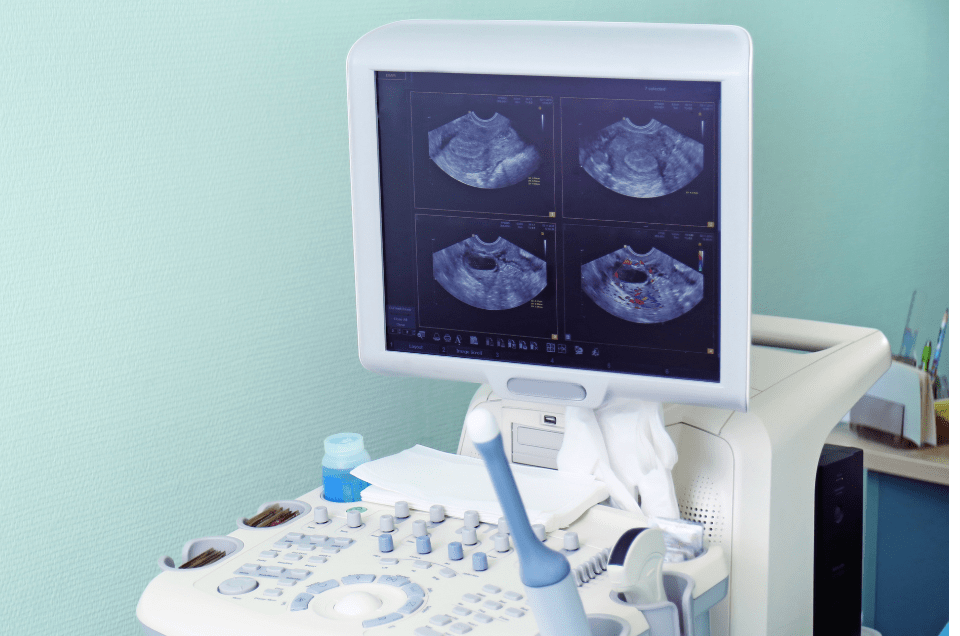
Transvaginal Ultrasound: Transvaginal ultrasound is a safe and painless procedure that is commonly used to examine the uterus and cervix. A small probe called a transducer is inserted into the vagina just like a tampon, allowing for detailed imaging of the pelvic organs. This technique helps identify the presence of cervical polyps and assess their size and location.
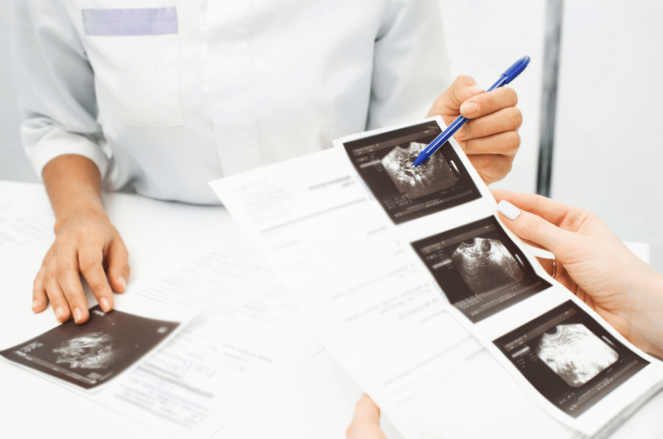
Differentiating from Other Conditions: Ultrasound images can help distinguish between cervical polyps and other growths, such as fibroids or tumors. This differentiation is crucial for proper treatment planning.
Monitoring: Ultrasound can be used to monitor the growth and changes of cervical polyps over time. This is particularly important for patients with more significant or recurrent polyps, as it helps healthcare providers make informed decisions regarding treatment options.
Examination To diagnose Cervical Polyp
Doctors can detect Polyps during a routine pelvic exam that appears like smooth, fingerlike growths on the cervix in red or purple color. Cervical polyps are of two types, ectocervical and endocervical. Ectocervical polyps form on the cervix’s outer surface layer of cells whereas endocervical polyps arise from the cervical glands.
Endocervical polyps are the most common type of cervical polyp. Postmenopausal women are prone to have ectocervical polyps and women who are premenopausal are prone to are more likely to develop endocervical polyps.
Biopsies, or tissue samples, are taken to the laboratory for testing. Typically, the results indicate benign polyp cells. In very rare cases, precancerous patterns known as neoplastic changes may be present.
Diagnosing cervical polyps involves a detailed examination process that encompasses medical history assessment, physical examination, and certain procedures like hysteroscopy, transvaginal ultrasound, and potentially biopsy. A good cooperation between the patient and healthcare provider is crucial to ensure accurate diagnosis, effective management, and timely treatment if required.